FIGURE 3.
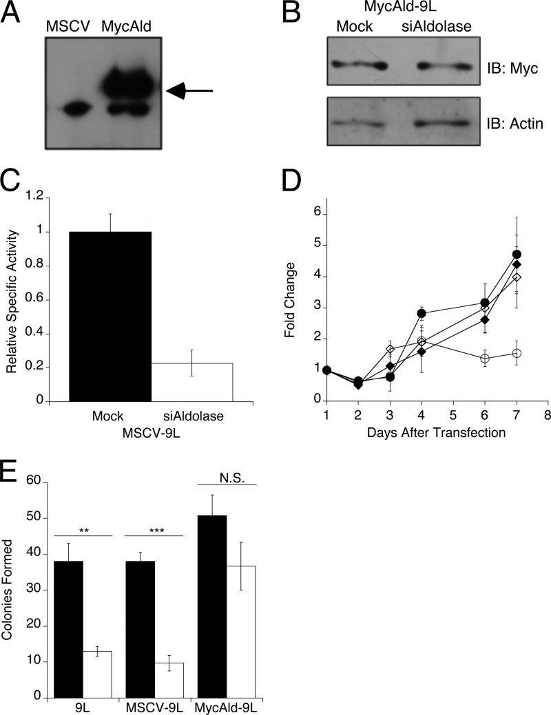
Rescue of 9L cells with exogenously expressed aldolase. A, anti-Myc immunoblot showing expression of Myc-tagged rabbit aldolase A in MycAld cells, but not in MSCV cells. Arrow indicates the position of 43 kDa, the expected size of the Myc-tagged aldolase. B, immunoblot showing Myc-tagged rabbit aldolase A expression in lysates following transfection of mock-treated and aldolase-knockdown (siAldolase) MycAld-9L cells. Cells were harvested 4 days after transfection, protein extracted, and assayed for MycAldolase via immunoblot. Actin was blotted as a loading control. C, MSCV-9L cells transfected with siRNAs to mouse aldolase A were monitored for knockdown by aldolase activity assay as described in Fig. 1A. Mock-transfected (0.02 ± 0.0014 units/mg) (black) and aldolase siRNA-transfected (white) values are shown. Error bars are represented as S.E. D, the number of cells/day were measured and plotted as described in Fig. 2 for mock (black) or siRNA to aldolase (open) transfected MSCV-9L (●, ○) and MycAld-9L (♦,♢) cells, respectively. Error bars are represented as 1 S.D. E, soft agar colony assay for parental 9L, MSCV-9L, or MycAld-9L mock-transfected (black) or aldolase-siRNA transfected (white) cells. After 14 days in soft agar, colonies were counted. Error bars represent 1 S.D. Statistical significance determined by t test is represented as ** (p < 0.005), triple asterisks (p < 0.001), or not significant (N.S., p > 0.05). IB, immunoblot.

