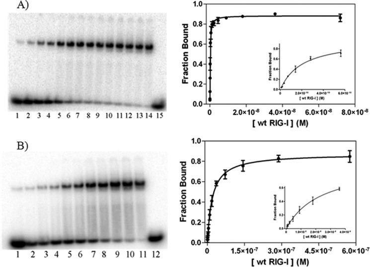
FIGURE 2.
Binding analysis of full-length RIG-I to 14 dsRNA substrates. A, EMSA of full-length RIG-I binding to 5′-ppp 14 dsRNA. Full-length RIG-I concentrations for lanes 1–7 are 9, 18, 35, 70, 140, 280, and 561 pm, respectively; concentrations for lanes 8–15 are 1, 2, 5, 9, 18, 36, 72, and 0 nm, respectively. The dsRNA concentration is 10 pm. The binding curve of full-length RIG-I to 5′-ppp 14 dsRNA is as follows: Kd = 0.159 ± 0.020 nm; R2 = 0.99. B, EMSA of full-length RIG-I binding to 5′-OH 14 dsRNA. Full-length RIG-I concentrations for lanes 1–12 are 0.5, 1, 2, 5, 9, 18, 36, 72, 140, 290, 580, and 0 nm, respectively. The dsRNA concentration is 1 nm. The binding curve of full-length RIG-I to 5′-OH 14 dsRNA is as follows: Kd = 20 ± 3 nm; R2 = 0.99. Insets, data for the six to seven lowest protein concentrations of the respective plots.

