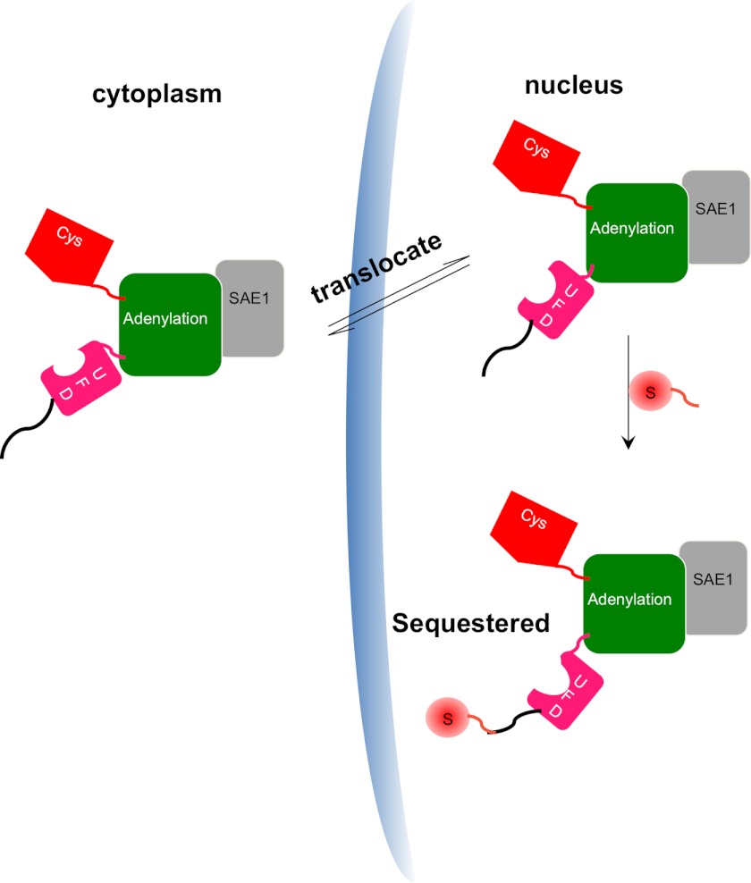
FIGURE 5.
A model for the role of SUMOylation in regulating SAE localization. The SAE1 subunit is shown in light gray, and the different domains of SAE2 are shown in color: the green domain contains the adenylation catalytic center; the red domain contains the catalytic Cys; and the pink region represents the ubiquitin-fold domain (UFD). SAE that is not SUMOylated at the SAE2 C terminus is rapidly shuttled in and out of nucleus, but SUMOylation of SAE2 at the C terminus sequesters the SAE in the nucleus.

