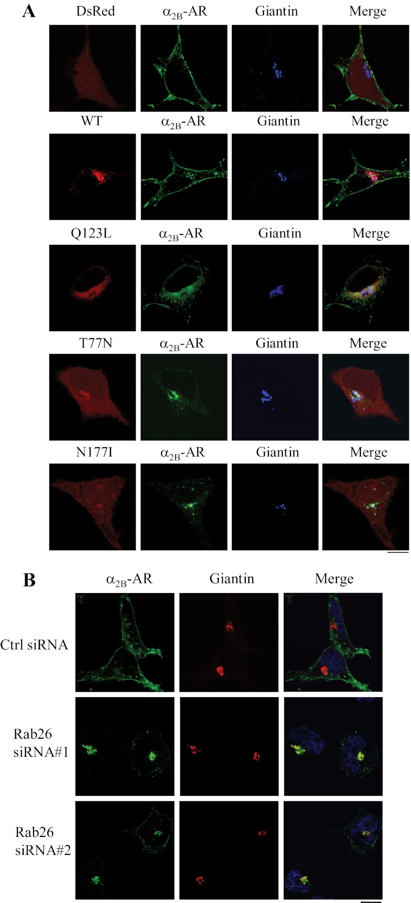
FIGURE 5.
Co-localization of α2B-AR with the Golgi marker giantin. A, co-localization of α2B-AR with giantin in cells expressing Rab26 or its mutants. HEK293 cells cultured on coverslips were transfected with α2B-AR-GFP together with the pDsRed-monomer-C1 vector, DsRed-tagged wild-type Rab26 (WT), or individual Rab26 mutant. Co-localization of α2B-AR with the Golgi marker giantin was revealed by confocal fluorescence microscopy following staining with giantin antibodies and Alexa Fluor 647-labeled secondary antibodies (1:300 dilution). Red, DsRed or DsRed-tagged Rab26; green, GFP-tagged α2B-AR; blue, giantin. B, co-localization of α2B-AR with giantin in cells expressing Rab26 siRNA. HEK293 cells were transfected with α2B-AR-GFP together with control siRNA or Rab26 siRNA. The cells were then stained with giantin antibodies and Alexa Fluor 594-labeled secondary antibodies. Green, GFP-tagged α2B-AR; red, giantin; yellow, co-localization of α2B-AR with giantin. In A and B, the data shown are representative images of at least three independent experiments. Scale bars, 10 μm.

