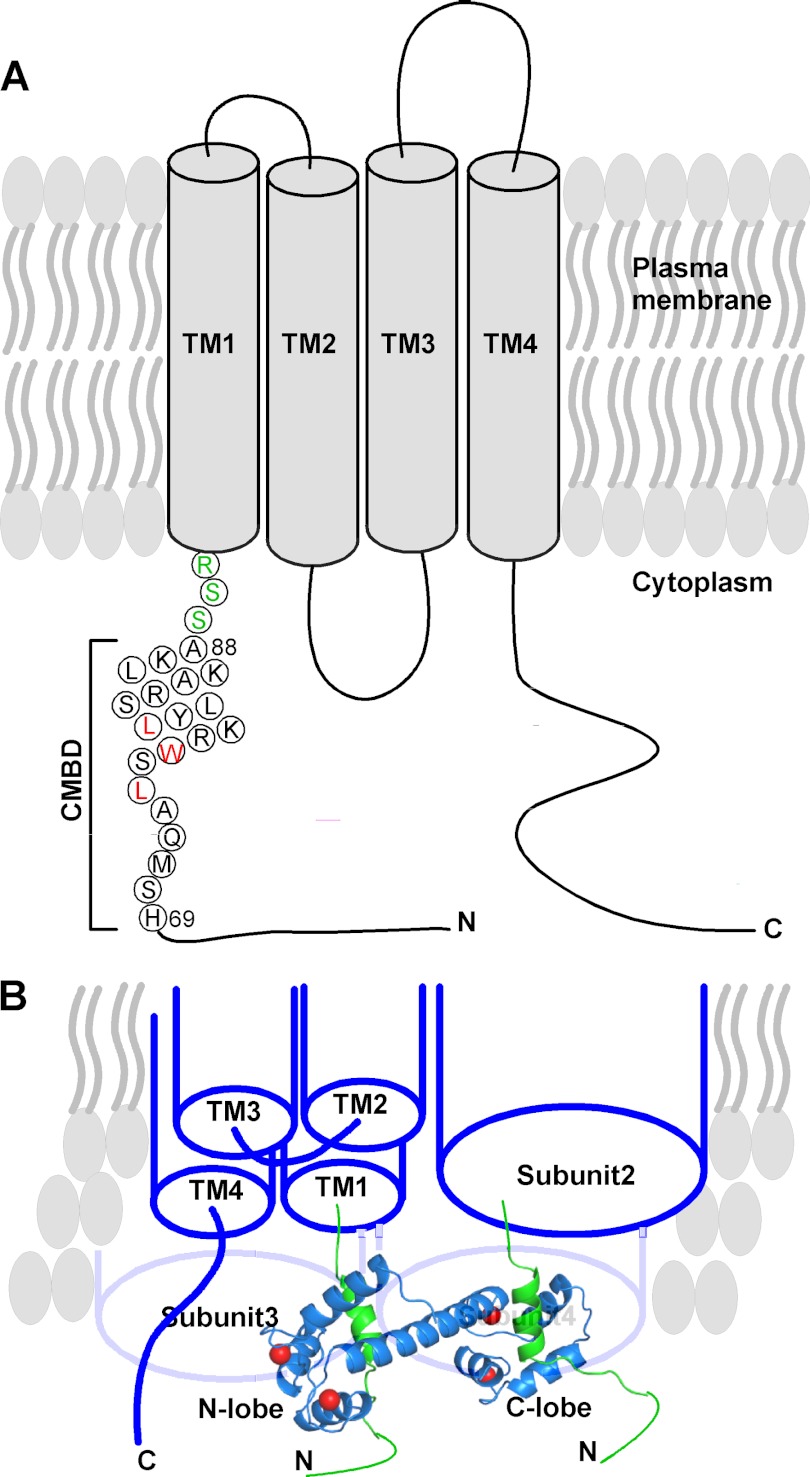
FIGURE 11.
Proposed mechanism for the interaction of CaM with Orai1 to carry out CDI. A, schematic presentation of the proposed Orai1 topology. The sequence of Orai1-CMBD from the structure is shown in circles. Leu74, Trp76, and Leu79 that are involved in hydrophobic interaction with CaM are colored in red. The three residues between the Orai1-CMBD and the first transmembrane helix (TM1) are colored in green. B, proposed binding of one CaM to two Orai1. Orai1 is shown as a tetramer. Subunit 1 is displayed with four transmembrane helices. The remaining three subunits are displayed as cylinders. Here, one CaM (blue ribbon) binds to two CMBDs (green ribbons) of the two adjacent Orai1 subunits, displacing STIM1 from binding to Orai1 to inactivate the channel. The other CaM for binding to subunits 3 and 4 is not displayed for clarity.

