Fig. 6.

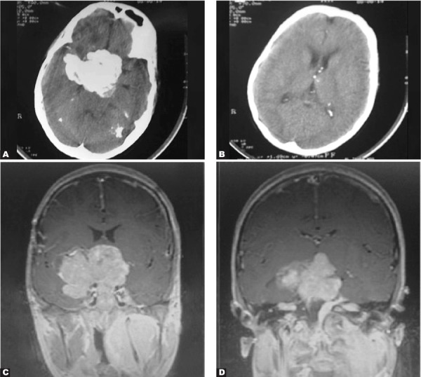
A 16-year-old boy with deafness and skull deformity. A and B. Axial post contrast CT scan C. Coronal post contrast T1-weighted image D.Coronal post Gd injection

A 16-year-old boy with deafness and skull deformity. A and B. Axial post contrast CT scan C. Coronal post contrast T1-weighted image D.Coronal post Gd injection