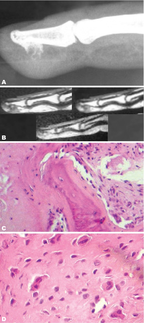
Fig. 4. A 45-year-old woman with swelling and pain in the distal phalanx of the right medius.

A. Lateral radiography of the right medius shows a calcified bone surface lesion developed from the palmar surface of the distal phalanx with soft tissue swelling but no adjacent bone abnormality. B. Sagittal MRI view of the right hand on T1W sequence, T1W sequence after intravenous Gadolinium injection and T2W sequence. The lesion shows a homogeneous low T1 signal and high T2 Signal with moderate enhancement after intravenous Gadolinium injection. C. Microscopic view (HE×200) showing bone trabecula associated with fibrous tissue. D. Microscopic view (HE×400) showing chondroid tissue made of chondrocytes of irregular size sometimes binucleated.
