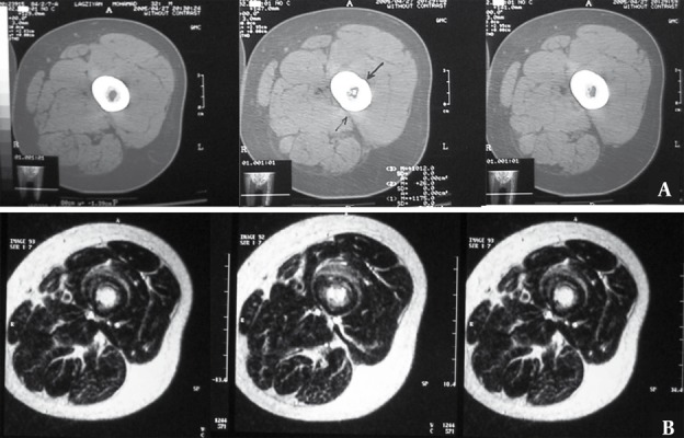
Figure 5. A 25-Year-Old Man Complaining of Pain in Mid FemurA.

A, CT scan through mid-femur. Nidus is visible in the medulary region only in one of the cuts.
B, MRI of the same patient reveals distinct hypersignal nidus in the medulary region in T2-W sequence in several cuts.
