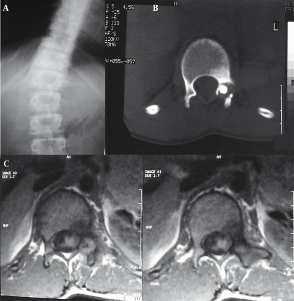
Figure 10. A 22-Year-Old Man With a Complaint of 5-Month Continuous Low Back Pain.

A, Radiograph revealed a sclerotic region in left pedicle of L1 vertebra along with scoliosis in thoracolumbar region.
B, Lumbar CT scan revealed osteolytic and osteoblastic lesion in pedicle of L1 vertebra.
C, MRI with T1 weighting demonstrated focal low signal intensity surrounded by soft tissue component replaced pedicle of L1 vertebra on the left side in the axial sections. Diffuse low signal intensity due to edema was seen in the adjacent bone.
