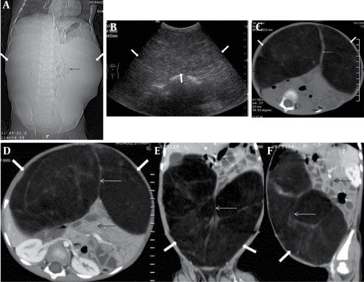
Figure 1. A 2.5-Year-Old Boy Presenting With Complaints of Gradual Abdominal Distention, Intermittent Abdominal Pain and Diarrhea.

A, Scannogram of the abdomen shows gross abdominal distension (thick white arrow) and centrally positioned bowel loops (thin black arrow).
B, Abdominal ultrasound in the same patient reveals a huge homogeneous echogenic mass (thick white arrow) occupying almost the entire abdomen.
C, (Plain) and D, E, F (Contrast-enhanced axial/coronal/sagittal) abdominal CT images of the same patient reveal a well-marginated non-enhancing intraperitoneal encapsulated mass (thick white arrow) of fat density (-70 to -140 HU) with few fibrous septae (thin white arrow) traversing the mass. Bowel loops and retroperitoneal structures are displaced posteriorly (thin black arrow). No inflammatory reaction or infiltration of surrounding tissues is noted.
