
Figure 2. A 26-year-old woman with swelling in the left mandible.
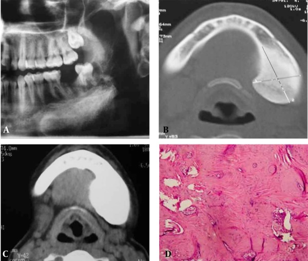
A, A moderately well-defined radiopaque lesion was seen adjacent to the left inferior border of the mandible; B, Axial CT showing a well-defined radiodense lesion on the left lingual aspect; C, CT with enhancement; D, Microscopically, the marrow spaces and lacunae of the mature bone were seen.