Figure 5.
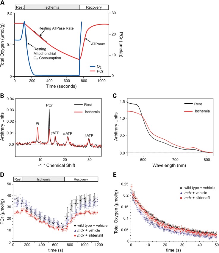
Phosphocreatine and oxygen fluxes in wild-type, untreated and sildenafil-treated mdx skeletal muscle. Schematic representation of the mild ischemia-recovery protocol used to measure mitochondrial oxidative phosphorylation function in vivo (A). Decreases in phosphocreatine (PCr) and O2 concentration were determined during mild hindlimb ischemia and used to calculate resting ATPase rates (P), a measure of resting mitochondria ATP synthesis, resting mitochondrial O2 consumption (O) and P/O coupling ratio. During muscle recovery and the resupply of O2 for oxidative phosphorylation, the rate of PCr recovery was used to calculate the maximal ATP synthesis rate (ATPmax) (B). Representative 31P spectra showing a decrease in PCr and proportional increase in inorganic phosphate (Pi) at the end of ischemia. ATP levels remain essentially constant since they are buffered by PCr (C). Representative optical spectra at rest of oxygenated hemoglobin (Hb) and myoglobin (Mb) and after ischemia of oxygen-desaturated Hb and Mb. These spectra were used to calculate muscle O2 concentration. Mean changes in PCr levels during ischemia–reperfusion in wild-type, mdx and sildenafil-treated mdx mice (D). Mean changes in O2 concentration during ischemia in wild-type, mdx and sildenafil-treated mdx mice (E). Number of mice per group: wild-type (7), mdx (6) and mdx + sildenafil (9).

