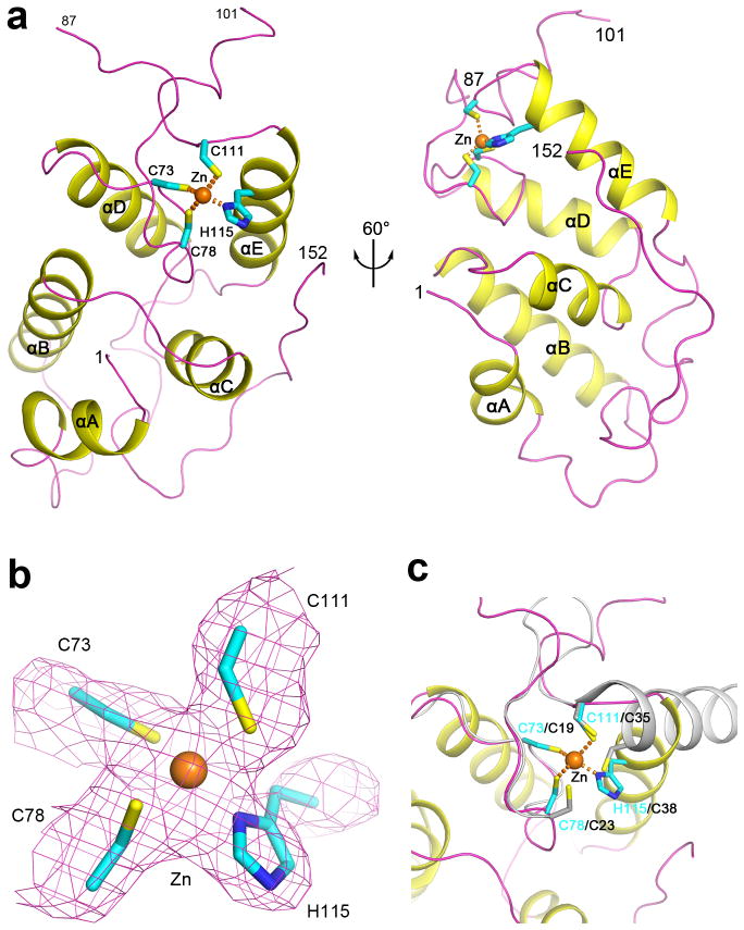
Figure 2.
Rtr1 is a new type of zinc finger protein. (a). Schematic drawing of the structure of K. lactis Rtr1. The zinc atom is shown as a sphere (in orange), and its four ligands are shown as stick models (in cyan). The two views are related by a rotation of ~60° around the vertical axis. (b). Simulated annealing omit Fo–Fc electron density for the zinc atom and its ligands in the structure of KlRtr1 at 2.5 Å resolution, contoured at 2.5σ. (c). Overlay of the zinc binding site of K. lactis Rtr1 (in color) and the zinc finger domain of Rabex-5 (in gray). The superposition is based on the first two zinc ligands. All the structure figures were produced with PyMOL (www.pymol.org).

