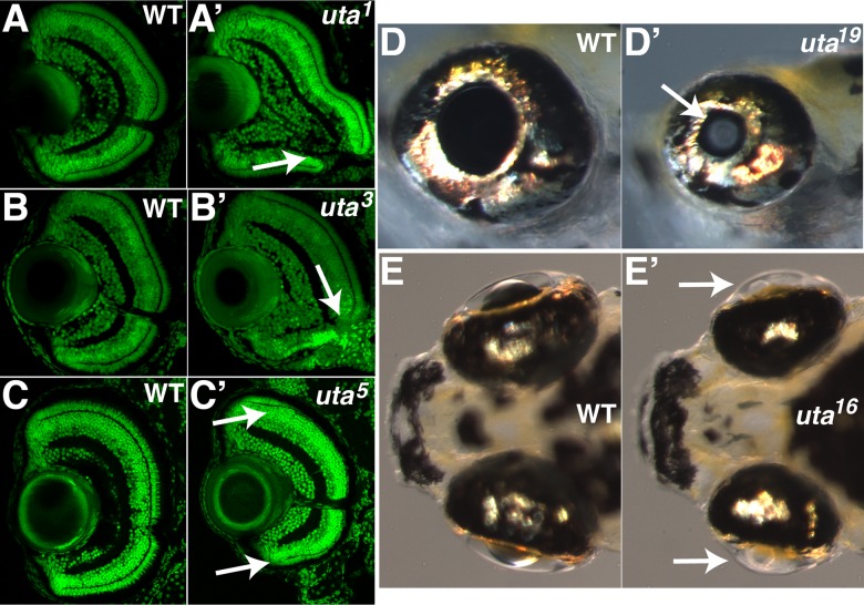
Figure 2. .
Examples of ocular mutants identified in the screen. uta1 (A') and uta3 mutants (B') display colobomas at 3 dpf when compared with phenotypically wild-type siblings ([A, B], respectively). Arrows in (A') and (B') point to the open choroid fissure in the mutants. uta5 mutants (C') lack photoreceptors in the central retina at 7 dpf when compared with phenotypically wild-type siblings (C). Photoreceptors are detected at the peripheral retina, adjacent to the ciliary marginal zones in mutant embryos (arrows in [C']). uta19 mutants (D') possess visible cataracts at 6 dpf when compared with phenotypically wild-type siblings (D). Arrow in (D') highlights cataract in mutant lens. uta16 mutants (E') possess severe defects in lens morphology at 7 dpf when compared with phenotypically wild-type siblings (E). Arrows in (E') highlight mutant lenses.

