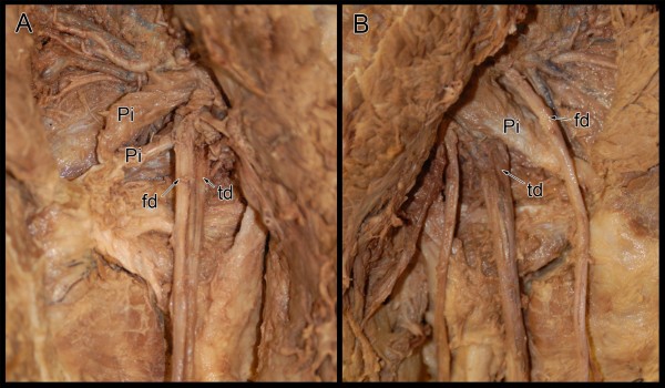
Figure 1.
Photograph reveals bilateral sciatic nerve variants on a single cadaveric specimen. The left side (A) revealed that sciatic nerve divided proximal to the piriformis muscle. The left common fibular division (fd) divided the piriformis (Pi) into two distinct bellies while the left tibial division (td) passed inferior and deep to the most caudal border of the piriformis muscle. On the right side (B), the sciatic nerve was split into the right common fibular (fd) and tibial (td) divisions proximal to the piriformis muscle (Pi). The right common fibular division passed superficial while the tibial division passed deep to the piriformis muscle.

