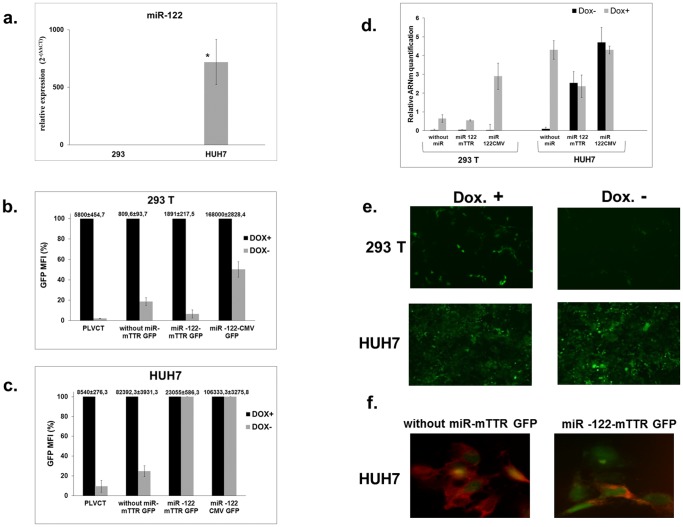
Figure 3. miR-122 concentrations and reporter gene expression in transduced cells.
(a) miR-122 expression levels in Huh7 and 293T cells detected by RT-qPCR. Three independent experiments were performed in duplicate. *Statistically significant differences P = 0.03 (two-tailed, unpaired Student’s t-test). (b) & (c) GFP expression in transduced Huh7 and 293T cells. After 10 days of culture, with or without doxycycline, transduced 293T (b) or Huh7 (c) cells were analyzed for GFP expression by FACS. Results are expressed as a percentage of MFI. In the TetR-KRAB system, the expression of GFP is optimal in the presence of doxycycline and corresponds to 100% of the MFI. The raw data corresponding to 100% are indicated above each histogram bar. (d) RT-qPCR of GFP mRNA normalized to values obtained for 18S RNA. Data shown are mean and error bars indicate the standard deviation (SD) of 3 independent experiments performed in triplicate. (e) Representative images of GFP expression in cells at 10 days after transduction. Lentiviral vectors encoding TetR-KRAB mRNA with four target sequences for miR-122 and GFP gene under the control of a liver specific promoter in 293T and Huh7 cells with or without doxycycline treatment. Magnifications ×50. (f) Representative high power fields showing immunofluorescence staining of TetR-KRAB (red) in Huh7 cells transduced with recombinant lentiviruses. Four target sequences for miR-122 were absent (left panel) or present (right panel) in the TetR-KRAB-expressing cassettes of the vectors, which also produced GFP (green) constitutively. Magnifications ×400.

