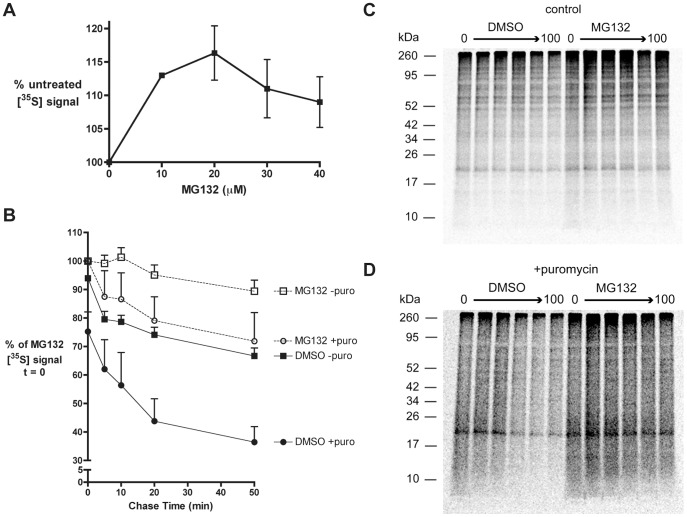
Figure 3. Treatment with puromycin increases the fraction of rapidly degraded polypeptides.
A. 293-Kb cells were labeled with [35S]-Met for 10 minutes in the presence of 0 to 40 µM MG132. [35S] incorporation was measured as in Fig. 1A and normalized to controls without proteasome inhibitor (n = 3; mean ± s.e.m.) B. 293-Kb cells were pulse labeled with [35S]-Met +/−20 µM puro and +/−20 µM MG132 for 5 minutes, then chased from 0 to 50 minutes in the presence of excess cold methionine, CHX and +/−20 µM MG132. DMSO is a solvent control for MG132. The chase was terminated at the indicated time points by the addition of TCA to cell suspensions to precipitate polypeptides. TCA precipitates were solubilized and [35S] was measured by liquid scintillation counting (n ≥4; mean ± s.e.m.) C and D. Solubilized TCA precipitates from cells radiolabeled in the absence (C) or presence (D) of 20 µM puro were separated by tricine SDS-PAGE on 10% gels. Gels were dried and exposed to a PhosphorImager plate overnight. Note that for D, the darkness of the image has been enhanced in order to see the contrast in degradation rates between C and D more clearly.

