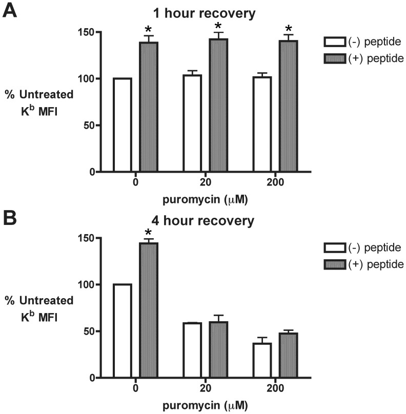
Figure 7. Time-dependent inhibition of MHC class I pathway function following puromycin treatment.
A and B. 293-Kb cells were stripped of cell surface MHC I peptides as in Fig. 5. Recovery of cell surface Kb was conducted in the presence of varying concentrations of puro for one (A) and four (B) hours. During the final 30 minutes of the recovery, cells were treated with either distilled water ((−) peptide) or 5 µM SIINFEKL peptide ((+) peptide) to promote the export of Kb to the cell surface [31]. Flow cytometry was used to measure total cell surface Kb and the MFI was normalized to untreated cells in the absence of exogenous SIINFEKL peptide (n = 3; mean ± s.e.m.; * p<0.05 for (−) peptide vs. (+) peptide).

