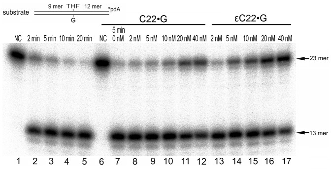
Figure 3. Inhibition of AP site cleavage activity of APE1 by C22•G and εC22•G oligonucleotide duplexes.
A solution of 1 nM of 3′-[32P]-labelled THF•G oligonucleotide duplex was incubated with 0.5 nM APE1 for 2–20 min at 37°C in the presence of increasing amounts of non-labelled 22 mer C22•G or εC22•G duplexes under NIR conditions. Lane 1, control, non-treated THF•G duplex; lane 2, as 1 but APE1 for 2 min; lane 3, as 2 but 5 min; lane 4, as 2 but 10 min; lane 5, as 2 but 20 min; Lane 6, as 1; lane 7, THF•G duplex with APE1 for 5 min; lane 8, as 7 but 2 nM C•G; lane 9, as 7 but 5 nM C•G; lane 10, as 7 but 10 nM C•G; lane 11, as 7 but 20 nM C•G; lane 12, as 7 but 40 nM C•G; lane 13, as 7 but 2 nM εC22•G; lane 14, as 7 but 5 nM εC22•G; lane 15, as 7 but 10 nM εC22•G; lane 16, as 7 but 20 nM εC22•G; lane 17, as 7 but 40 nM εC22•G. For details see Materials and Methods.

