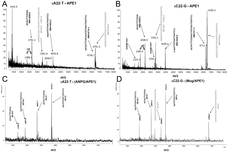
Figure 4. MALDI-TOF MS analysis of the mixture of oligonucleotides arising from the incubation of the 22 mer oligonucleotide duplexes containing a single εA or εC residue with APE1 or DNA glycosylases.
Typically, a solution of 10 pmol of the lesion containing oligonucleotide duplexes was incubated with either 10 nM APE1 under NIR conditions at 37°C for 17 h or 50 nM ANPG or 50 nM MUG at 37°C for 30 min and subsequently with 10 nM APE1 at 37°C for 30 min under “BER+Mg2+” reaction conditions. (A) Treatment of εA22•T duplex with APE1; (B) Treatment of εC22•G duplex with APE1; (C) Treatment of εA22•T duplex with ANPG and APE1; (D) Treatment of εC22•G duplex with MUG and APE1. Peaks corresponding to complementary strands are indicated in grey. For details see Materials and Methods.

