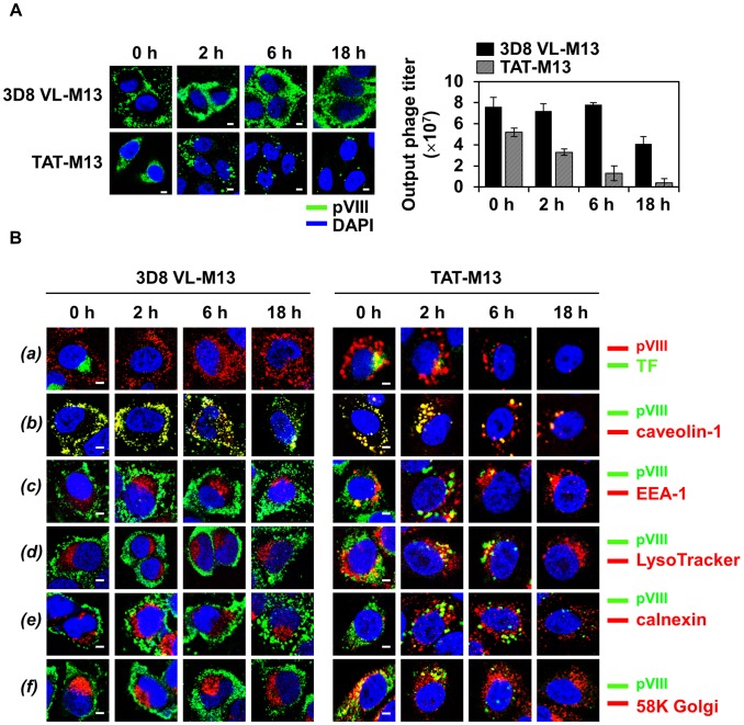
Figure 5. Internalized 3D8 VL-M13 phage routes to the cytosol and remains stable without further trafficking to other subcellular compartments, whereas TAT-M13 phage is routed to other subcellular compartments before rapid degradation in the lysosome.
In the following pulse-chase experiments, HeLa cells (1×10⁶ cells) in serum-free medium were treated at 37°C with 1012 CFU of 3D8 VL-M13 for 2 h or 1013 CFU of TAT-M13 for 30 min. Then surface bound phages were removed by multiple washes with low pH glycine buffer and then internalized phages were chased at 0, 2, 6, and 18 h. (A) Time-course intracellular localization of internalized phages was visualized by confocal immunofluorescence microscopy or time-course output phages were quantified by the CFU assay and represented as mean ± S.E. (error bars) of three independent experiments. (B) Time-course intracellular trafficking of internalized phages monitored by co-localization with transferrin (TF, a), caveolin-1 (b), early endosome marker EEA-1 (c), late endosome/lysosome tracker LysoTracker (d), ER marker calnexin (e), or Golgi marker 58K Golgi protein (f), as visualized by confocal immunofluorescence microscopy. In (A) and (B), internalized phages were visualized by confocal immunofluorescence microscopy with primary anti-pVIII antibody and secondary FITC-anti-mouse antibody (A and B, a, green) or TRITC-anti-mouse antibody (B, b-f, green). In (A) and (B), magnification, ×400; scale bar, 5 µm.

