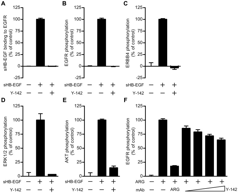
Figure 3. Neutralizing activities of Y-142 against sHB-EGF and ARG signaling.
(A) Inhibitory activity of Y-142 to sHB-EGF binding to EGFR. EGFR-hFc was incubated in an anti-human IgG Fc antibody-coated plate. Y-142 was then incubated at a concentration of 6.7 nM in the presence of 0.63 nM biotinylated sHB-EGF for 1 hour at 37°C. sHB-EGF bound to EGFR-hFc was detected by HRP-labeled streptavidin. sHB-EGF binding to EGFR-hFc in the presence of Y-142 was calculated as a percentage of the “control” sHB-EGF binding to EGFR which occurred without Y-142. Data points represent the mean + SD of values acquired in triplicate. (B) Neutralizing activity of Y-142 against EGFR phosphorylation. SK-OV-3 cells were treated with 10 nM sHB-EGF and 67 nM Y-142. Cell lysates were incubated in an anti-EGFR antibody-coated plate, followed by an incubation with HRP-labeled anti-phosphorytosine antibody. EGFR phosphorylation in the presence of Y-142 was calculated as a percentage of the “control” EGFR phosphorylation which occurred without Y-142. Data points represent the mean + SD of values acquired in triplicate. (C) Neutralizing activity of Y-142 against ERBB4 phosphorylation. Cell lysates of T47D cells as prepared in Fig. 3A were incubated on an anti-ERBB4 antibody-coated plate. The phosphorylation of ERBB4 was detected by a sulfo-tagged anti-phosphotyrosine antibody. ERBB4 phosphorylation in the presence of Y-142 was calculated as a percentage of the “control” ERBB4 phosphorylation which occurred without Y-142. Data points represent the mean + SD of values acquired in duplicate. (D) and (E) Neutralizing activity of Y-142 against (D) ERK1/2 phosphorylation and (E) AKT phosphorylation. In (D) and (E) SK-OV-3 cells treated with 10 nM sHB-EGF and 200 nM Y-142 were stained with an anti-phosphorylated ERK1/2 antibody or an anti-phosphorylated AKT antibody, respectively, followed by an Alexa488-labeled anti-rabbit IgG antibody. Phosphorylated ERK1/2 and phosphorylated AKT were both detected with an ImageXpress Micro instrument and calculated as a percentage of the “control” phosphorylation levels which occurred without Y-142. Data points represent the mean + SD of values acquired in duplicate. (F) Neutralizing activity of Y-142 to ARG. SK-OV-3 cells were treated with 10 nM ARG plus various concentrations of Y-142 (2 nM, 6.7 nM, 20 nM, and 67 nM). Anti-ARG monoclonal antibody (67 nM) was used as a positive control. Cell lysates were incubated in an anti-EGFR antibody-coated plate followed by an incubation with an HRP-labeled anti-phosphorytosine antibody. EGFR phosphorylation was calculated as a percentage of the “control” EGFR phosphorylation which occurred without Y-142. Data points represent the mean + SD of values acquired in triplicate.

