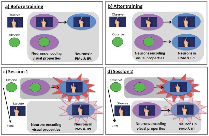
Figure 2. Schematic representation of the fMRI adaptation logic.
Note that although reference is made to mirror neurons, fMRI data is driven by populations of neurons. Purple ovals denote populations of sensory neurons encoding visual properties of stimuli; blue ovals denote populations of motor neurons responsible for action execution. a) Before training, motor neurons are activated by observation of actions (top) but not by observation of shapes (bottom). These cells are therefore mirror neurons b) Training where participants respond to each arbitrary geometric shape with a distinctive action establishes novel excitatory links (broken arrow) between neurons encoding sensory properties of each shapes and motor neurons encoding the trained action. c) Session 1. Adaptation from shape observation to action execution (signified by paler flash on right), and vice versa, shows that, as a result of training, the shapes activate neuronal populations with motor properties. d) Session 2. Adaptation from shape observation to action observation, and vice versa, shows that shape and action observation activates common neuronal populations; i.e. cells with mirror properties. Session 2 adaptation would not have occurred if experimental training had linked visual neurons with i) purely motor neurons, ii) canonical neurons, or iii) logically related mirror neurons. The training must have linked neurons encoding the sensory properties of the geometric shapes with neurons that were already encoding both sensory and motor properties of action, i.e. congruent mirror neurons.

