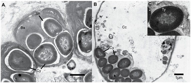
Figure 2. Transmission electron microscopy of cyanobionts from the sponge Hymeniacidon perlevis (Sponge ID HYM5B).
(A) Xenococcus like morphotypes observed at dividing stage with prominent baeocytes with gelatinous outer sheath. (B) Cyanobacterial colony showing compactly packed spiral thylakoids in choanocyte chamber. (C) Insight showing zoomed in image of cyanobacterial cell. (Cy, Cyanobacteria; Ba, Baeocytes; Sh, Sheath; Cc, choanocyte chamber; Ty, Thylakoid).

