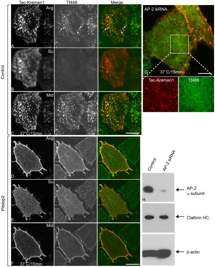
Figure 3. Inhibition of endocytosis of Tac-Kremen1 with CME inhibitors.
HeLa cells were transfected with Tac-Kremen1 and then were starved for 45 min in DMEM, 0.1% FBS at 37°C. Starved cells were mixed with 0.1% DMSO (final) (A-C) or 30 µM pitstop2 (final) (D-F) for 15 min at 37°C. Mouse monoclonal anti-Tac and transferrin-Alexa488 (Tf488) were added, and cells were incubated for 15 min at 37°C. Cells were fixed, permeabilized, and stained for confocal microscopy. The Z stack data were stitched by FIJI. (A and D) Representatives of averaged images. (B and E) Single optical sections of surface localization. (C and F) Single optical section of mid sections. (G) HeLa cells were depleted of AP-2 α-subunit by treatment with siRNA and then were transfected with Tac-Kremen1. Cells were starved in DMEM, 0.1% BSA, and 10 mM Hepes (pH 7.5) before incubation on ice with mouse monoclonal anti-Tac and transferrin-Alexa488 for 1 h. The endocytosis assay was performed by shifting the temperature to 37°C for 15 min. Cells were immediately fixed, permeabilized, incubated with anti-mouse-IgG-Cy3 and visualized by confocal microscopy. Scale bar 10 µm. (H) Immunoblot confirming the knockdown of AP-2 α-subunit; levels of clathrin-HC remained unchanged. β-actin was used as a loading control.

