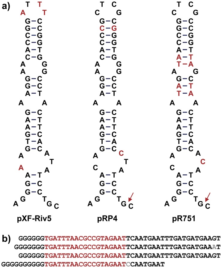
Figure 3. Structure of oriT and oriV.
A) oriT inverted repeats from pXF-RIV5, pRP4, and pR751 are shown as stem-loop structures to emphasize potential base parings. Unique bases are shown in red; bases conserved in at least two structures are shown in black. Red arrows indicate experimentally determined cleavage sites in pRP4 and pR751 [21]. B) Tandem repeats in pXF-RIV5 oriV are aligned and the 19-bp core is shown in red. Residues that vary from the consensus are shown in gray. Nucleotides shown (34333 – 34503) are contiguous.

