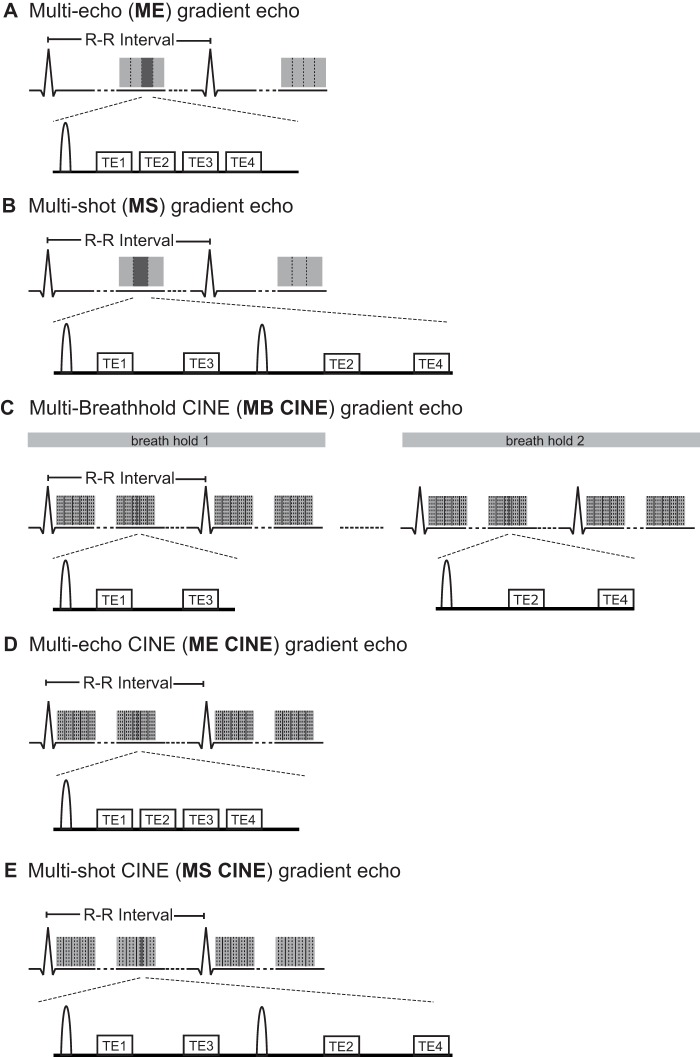
Figure 1. Synopsis of multi-echo gradient echo strategies used for T2 * mapping at 7.0 T.
A). Conventional multi-echo (ME) gradient echo for single cardiac phase myocardial T2 * mapping. Multiple echoes are acquired after excitation to obtain a set of T2 * weighted images. The competing constraints of inter echo time and spatial resolution inherent to the ME approach are addressed by the B) interleaved multi-shot multi-echo (MS) gradient echo technique. In MS a set of excitations is employed together with echo interleaving echoes to acquire a set of T2 * weighted images. C) The multi-breath-hold multi-echo (MB CINE) gradient echo technique allows myocardial CINE T2 * mapping by interleaving the echoes over several breath-holds. For benchmarking D) multi-echo CINE (ME CINE) gradient echo and E) multi-shot multi-echo CINE (MS CINE) were applied for T2 * mapping in phantom studies. To guide the eye vertical dashed lines refer to k-space lines. Vertical solid lines refer to cardiac phases. A unipolar readout using gradient flyback was applied for all strategies.

