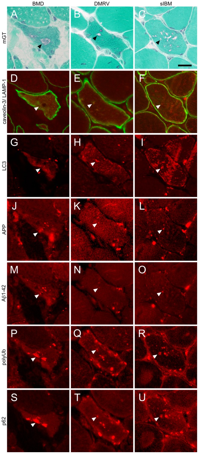
Figure 2. Immunohistochemical characteristics of RV in BMD compared to DMRV and sIBM.

Representative transverse serial sections of biopsied skeletal muscles from BMD with RV (left column), DMRV (center column) and sIBM (right column) patients. A–C: mGT staining similarly highlights the fibers with RVs (arrowheads) in all patients. D–F: LAMP-1 (red) co-stained with caveolin-3 (green), G–I: LC3, J–L: APP, M–O: Aβ1-42, P–R: polyUb proteins, and S–U: p62. Immunofluorescent signals are observed around RVs (arrowhead). Scale bar: 25 µm.
