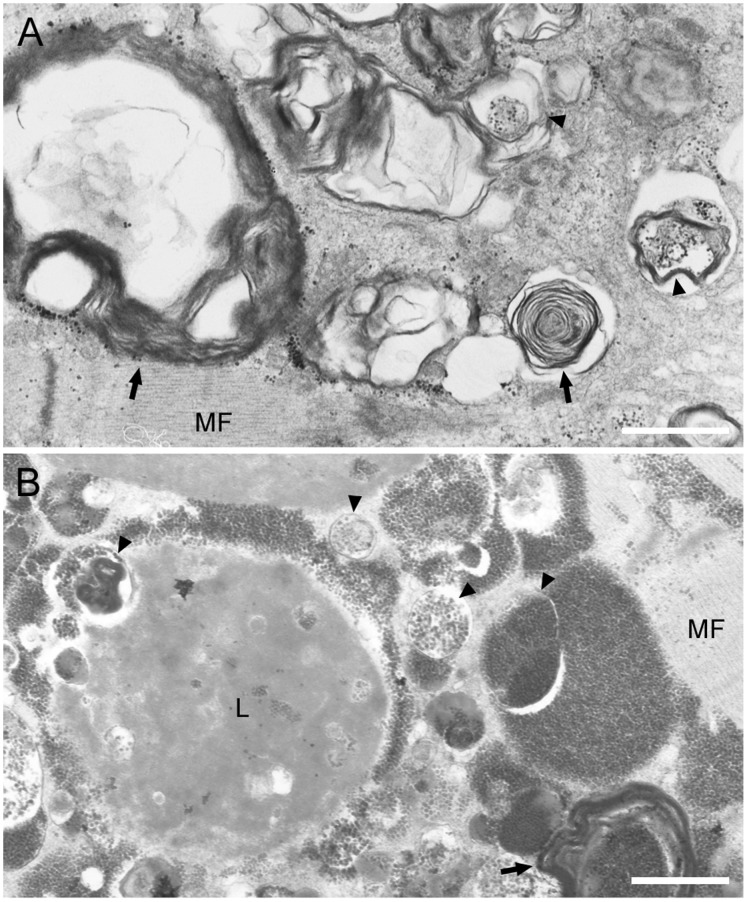
Figure 4. Areas of RVs in BMD myofibers show typical electron microscopic characteristics of autophagic vacuoles. A:
Accumulation of autophagic vacuoles (arrowheads), various cellular debris, and multilamellar bodies (arrow) are seen in myofibers of some BMD patients. Note the intact arrangement of myofibrils (MF) surrounding autophagic area. B: In areas with or without autophagy, lipofuscin deposit (L) is seen. Scale bars: 1 µm.

