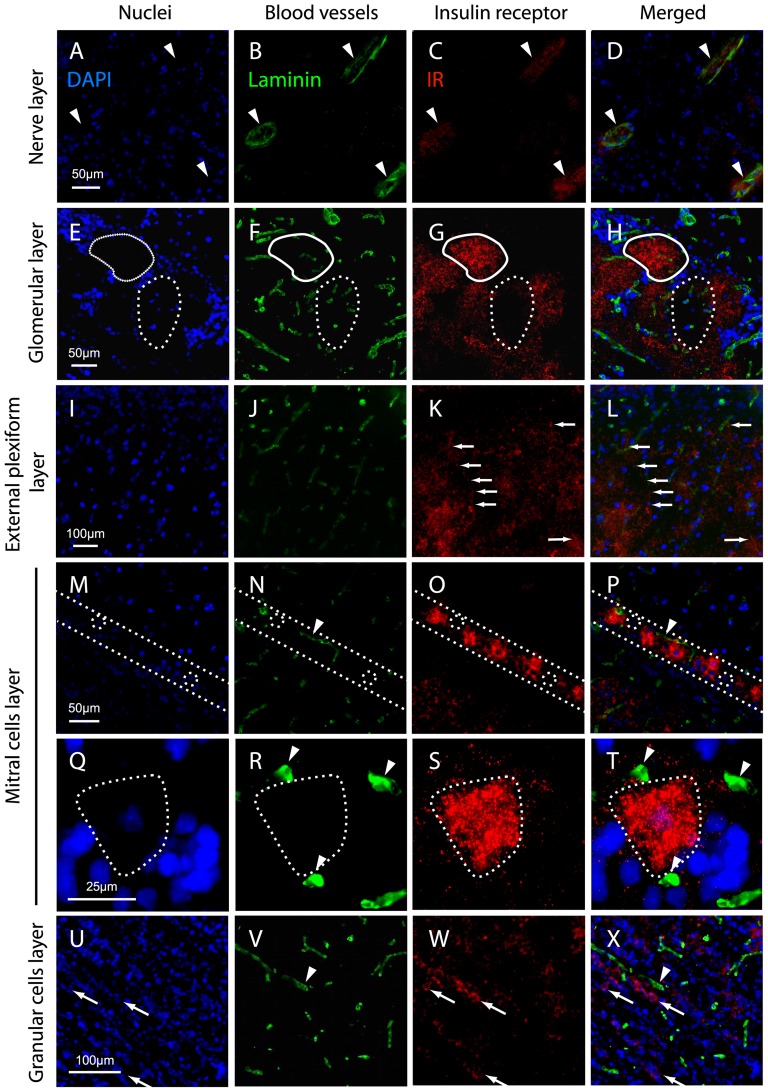
Figure 8. IR localization in the OB layers.
Representative images of the various layers of the main OB of adult rats obtained from frontal frozen sections (14 µm) stained with the nuclear marker DAPI (A, E, I, M, Q, U) and with double immunostaining of the blood vessel marker laminin (B, F, J, N, R, V) and IR (C, G, K, O, S, W). The signals were visualized on an epifluorescence microscope using sequential channel scanning with a merged overlay (D, H, L, P, T, X). In the nerve layer (A–D), IR immunostaining is restricted to the endothelium of blood vessels (white arrowheads). In the glomerular layer (E–H), IR immunostaining is abundant is the neuropil of some glomeruli (full line). Some glomeruli are not labeled (dotted line). In the external plexiform layer (I–L), a punctiform IR immunostaining is scattered on labeled fibers (arrows). In the mitral cell layer (M–P), between the dotted lines, IR immunostaining is present in most cell bodies, although some mitral cells are not labeled (dotted circle). The labeled cell bodies are consistently surrounded by blood microvessels (arrowhead). (Q–T) Enlargement of a mitral cell body (dotted line) surrounded by three blood microvessels (arrowheads). In the granular cell layer (U–X), IR immunostaining is located in clusters of labeled granular cell bodies (arrows). The labeled clusters are often located close to blood microvessels (arrowhead).

