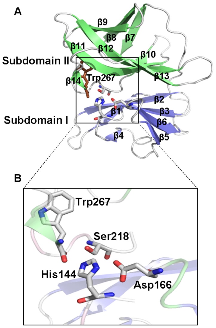
Figure 1. Structure of Aura Virus Capsid Protease.
(A) Overall structure of the capsid protein with two β-barrels of subdomains I and II colored in blue and green respectively. The catalytic triad residues and Trp267 are shown in sticks; (B) Close-up view of the active site shows catalytic triad composed of Ser218, Asp166 and His144 along with the carboxy-terminal Trp267 approaching the active site.

