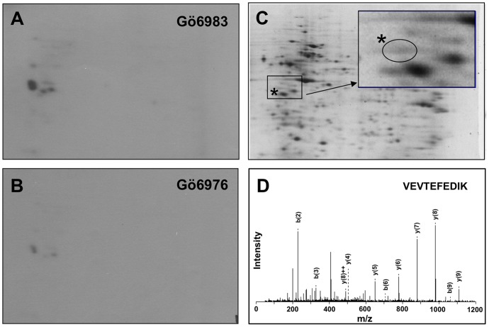
Figure 1. SET protein in Jurkat nuclear extract is phosphorylated by PKD2.
Activated PKD2 was immunoprecipitated from lysate of Jurkat cells stimulated with anti-CD3ε antibody. The heat-inactivated (50°C, 5 min) Jurkat nuclear extract was used as a substrate for PKD2 and analyzed by 2D-gel electrophoresis. (A) Activated PKD2 was incubated with the nuclear extracts in the presence of [γ-32P]-ATP and Gö6983 (an irrelevant PKC inhibitor) and the radioactivity of 2D-electrophregram was detected. (B) Activated PKD2 was incubated with the nuclear extracts in the presence of [γ-32P]-ATP and Gö6976 (PKD2 inhibitor) and the radioactivity of 2D-electrophoregram was detected. (C) The proteins in the Jurkat nuclear extract treated with non-radioactive ATP and PKD2 were stained with syproruby. Inset; the enlarged view of the boxed area. A protein spot indicated by the asterisks, which overlaps to the radioactive spots indicated in A or in B was analyzed by mass spectrometry. (D) One of the MS/MS spectra assigned to be SET protein is shown (m/z = 604, 110VEVTEFEDIK119).

