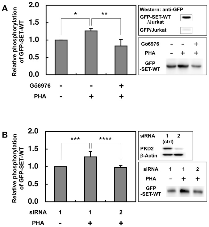
Figure 2. PKD2 is involved in the up-regulation of SET phoshorylation in activated Jurkat cells.
(A) After Jurkat cells expressing GFP-SET were stimulated by PHA (2 µg/ml) with or without Gö6976 (3 µM), the phosphorylated GFP-tagged SET was collected by PhosphoProtein Purification kit and separated by SDS-PAGE. The phosphoprotein was transferred onto the PVDF membrane and quantified by ECL using anti-GFP antibody and the secondary antibody. The typical blot was shown in the lower-right panel. The luminescence of samples from cells without both stimulation and the inhibitor was assigned to be 1. The results were expressed as mean +SD for three independent experiments. *, p<0.01; **, p<0.05. As shown in the upper-right panel, phosphorylated GFP was not detected from the eluate of the PHA stimulated Jurkat cells expressing GFP only. (B) Jurkat cells expressing GFP-SET treated with control (ctrl) siRNA1 had no effect on the PKD2 expression, while siRNA2 markedly reduced the PKD2 expression (upper-right panel). The typical blot was shown in the lower-right panel. The luminescence of the samples from non-stimulated and siRNA1-treated cells was assigned to be 1. The results were expressed as mean +SD for three independent experiments. ***, p<0.05; ****, p<0.05, as determined by Student's t test.

