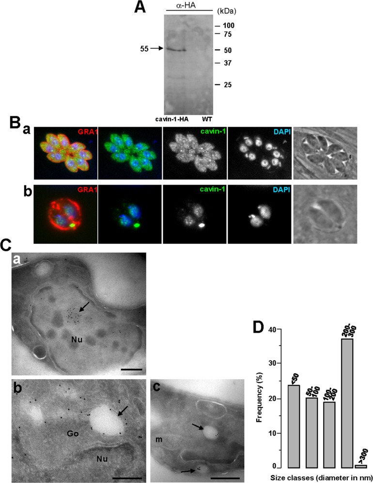
Figure 6. Expression and localization of cavin-1 in Toxoplasma.
(A) Expression of cavin-1-HA by transgenic parasites. Immunoblots of homogenates from wild-type T. gondii (left lane) and Toxoplasma stably expressing cavin-1-HA (right lane) revealed with anti-HA antibodies (α-HA). The Western blot shows a band at ∼55-kDa in transgenic parasites. (B) Localization of cavin-1-HA in transgenic parasites. IFA assays of cavin-1-HA-expressing Toxoplasma using anti-HA antibodies showing staining in vesicles (panels a and b), the cytosol (panel a) and the nucleus (DAPI; panel b). (C) Ultrastructure of cavin-1-containing structures formed in transgenic Toxoplasma. ImmunoEM staining of intracellular Toxoplasma expressing cavin-1-HA using anti-HA antibodies revealed by 10 nm-protein A-gold particles confirming a staining within the nucleus (panel a), the cytosol (panel b) or on vesicles in the Golgi area (Go) (arrow in panel b) and vesicles randomly distributed in the cytoplasm (arrows in panel c). Arrow shows a membranous structure connecting two vesicles. m, mitochondrion; Nu, nucleus. Bars are 200. (D) Size distribution of cavin-1-containing vesicles labeled with anti-HA antibodies-protein A gold particles. The diameters in nm of over 70 gold-labeled vesicles were measured from 50 parasite sections taken at 66,000× magnification, and their frequency was tabulated.

