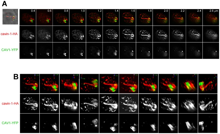
Figure 11. Interaction of CAV1-labeled structures with cavin-1-induced tubules in transgenic parasites.
Fluorescence microscopy of transgenic parasites co-expressing CAV1 and cavin-1. (A) A series of z-slices of two co-expressing transgenic parasites. Shown are phase contrast images overlaid with the corresponding fluorescence signals, merged images displaying CAV1 and cavin-1 fluorescence and images with only CAV1 or cavin-1 fluorescence shown. (B) 3D-reconstructions of z-stacks from the transgenic parasites in (A) shown in multiple rotated views.

