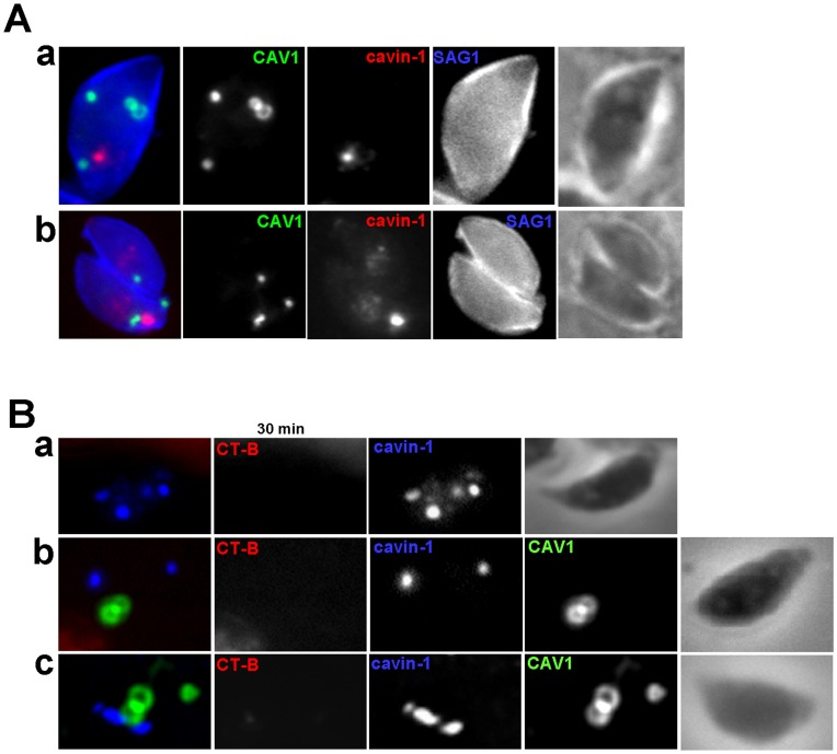
Figure 13. Assessment of caveolar endocytosis by CAV1-and/or cavin-1-expressing parasites.
(A) IFA of Toxoplasma coexpressing CAV1-YFP cavin-1-HA immunolabeled with antibodies against HA (cavin-1-HA) and SAG1 (plasma membrane, blue) showing no overlap with the plasma membrane. (B) Assessment of Alexa Fluor 594- CT-B distribution after 30 min-pulse by fluorescence microscopy in cavin-1-HA-expressing parasites (panel a) and CAV1-YFP- and cavin-1-HA-expressing Toxoplasma (panels b and c), showing no internalization of the CT-B regardless of the proximity of the structures containing CAV1 or cavin-1.

