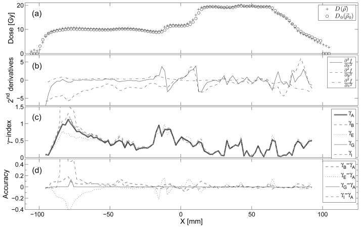
Figure 5.
(a) IMRT dose distributions along the X direction at Y = −31.8 mm and Z = 10 mm. (b) Second derivatives of the dose difference function defined by Eq. 3 with respect to the unit-less spatial coordinates, (c) γ (ρ⃗0) by methods defined in tab. 1. (d) Accuracy of the γ-index methods with respect to γA · γB shows the magnitude of the error in individual voxels (~ ± 0.1) caused by linear interpolation alone. γE and γI are more accurate than γB except in regions where the 2nd derivatives and γ are large. γG is the most accurate of the four methods compared to γA.

