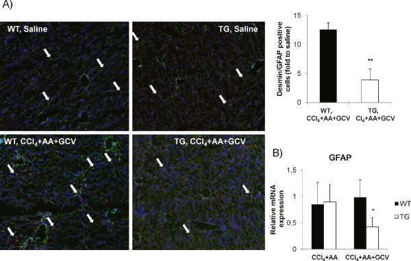
Figure 2. Validation of HSC depletion in vivo in TG mice treated with CCl4+AA+GCV.
(A) Reduction of desmin (green) and GFAP (red) positive cells, and merged images (yellow) by 65% (p<0.01) in TG mice treated with CCl4+AA+GCV, compared to WT mice as assessed by dual-immunofluorescence. (B) Relative GFAP mRNA expression (by qPCR) demonstrates a significant reduction in TG mice undergoing HSC depletion. Data are mean values from three independent experiments normalized to saline-treated mice. GCV treatment (without CCl4+AA) did not differ from saline treated group. Original magnification ×100. *p<0.05.

