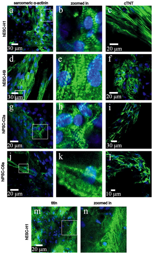
Figure 3. Immunostained cardiomyocytes derived from hESCs and hiPSCs.
(a–l), representative images of immunostained cardiac troponin T (TnT) and sarcomeric α-actinin in all four cell-lines at day 40. Striated staining patterns indicative of sarcomeres are seen in all hESC and hiPSC lines; however striations are most extensive in H1 cells, wherein titin immunostaining (m–n) reveals a level or sarcomeric networking that was not observed in the other cell-lines.

