
Figure 5. Different hyperscanning devices for hemodynamic and electric modalities.
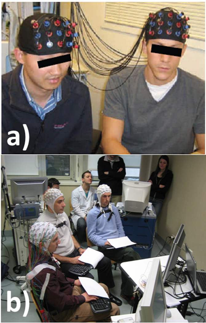
The figure shows in the upper part A) the split of the fibers to generate an NIRS hyperscanning device from one single NIRS device; in the lower part B) the EEG hyperscanning setup during an iterated Prisoner’s Dilemma task involving three subjects. Figures from Cui et al., 2011 and Astolfi et al., 2011.