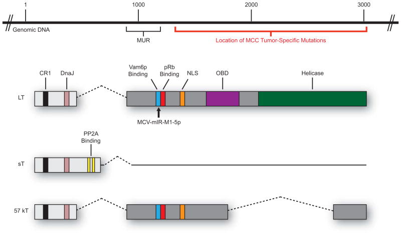
Figure 3. The MCPyV T antigen locus.
The MCPyV T antigen locus is located in the early region of the genome and encodes three T antigen isoforms that arise by alternative splicing. The large T antigen (LT) shares many traditional domains found in other polyomaviruses: CR1 (LXXLL; black), DnaJ (HPDKGG; pink), pRb-binding (LXCXE; red), a nuclear localization signal (NLS; orange), origin-binding domain (OBD; purple), and a helicase domain (green). LT also contains unique features, such as a binding site for Vam6p (blue) and a 200-amino acid “MCV-Unique Region” (MUR). The location of reported tumor-specific mutations in the 3′ region of LT is depicted with a red bar. The site of MCPyV microRNA (MCV-mIR-M1-5p) complementarity in LT is denoted (black arrow). The small t antigen (sT) shares the first exon with LT, but due to alternative splicing also contains a protein phosphatase 2A-binding site (PP2A; yellow). The 57 kT antigen is identical to LT up to the NLS motif, at which point differential splicing precludes the inclusion of complete OBD and helicase domains.

