Fig. 1.
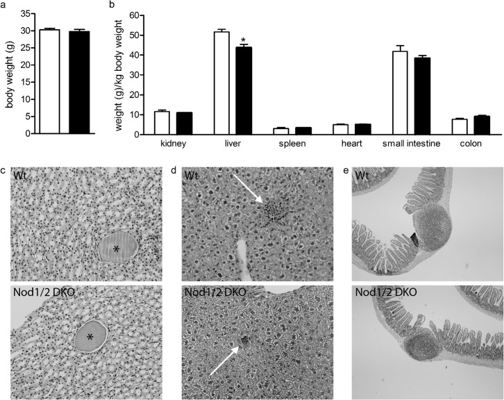
Body (a) and organ (b) weight of Wt (white bars) and Nod1/2 DKO (black bars) mice at the age of 3 months revealed no differences except for liver weight, which was lower in Nod1/2 DKO compared with Wt mice. (c) PasD-stained kidney sections (magnification 20×) of Wt (upper) and Nod1/2 DKO (lower) mice showing small cysts (asterisk). (d) HE-stained liver sections (magnification 20×) of Wt (upper) and Nod1/2 DKO (lower) mice showing small inflammatory infiltrates (white arrow). (e) HE-stained small intestinal sections (magnification 4×) of Wt (upper) and Nod1/2 DKO (lower) mice including Peyer's patches. Data are expressed as mean ± sem, n = 7. *P<0.05 compared with Wt.

