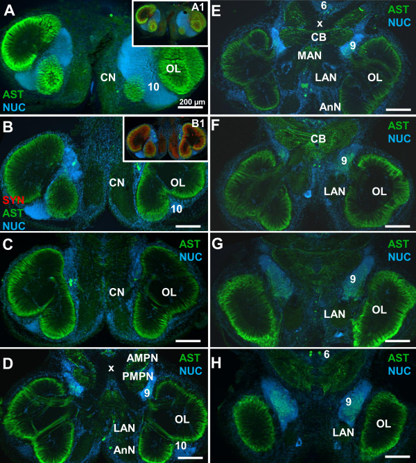
Figure 2.
A- type allatostatin-like immunoreactivity (ASTir) in the brain of C. clypeatus, low power views (conventional fluorescence with Apotome structured illumination) of a dorsal (A) to ventral (H) series of sections double labeled for ASTir (green) and the nuclear marker (blue). Insets A1 and B1 show additional labelling for synapsin (red). Abbreviations: Numbers 6, 9, 10 identify cell clusters, AMPN anterior medial protocerebral neuropil, AnN antenna 2 neuropil, CB central body, CN columnar neuropil, LAN lateral antenna 1 neuropil, OL olfactory lobe, PMPN posterior median protocerebral neuropil, x chiasm of the olfactory globular tract. The scale bar is 200 μm in all images.

