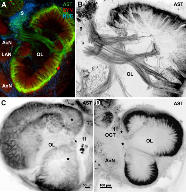
Figure 5.
Details of ASTir in the olfactory lobes (OL) as seen in conventional fluorescence with Apotome structured illumination (A) as well as confocal laser scanning microscopy (B-D). A shows the olfactory lobe triple immunolabeled for ASTir (green), synapsin (red) and the nuclear marker (blue). B-D the allatostatin channel only (images are black/white inverted), showing the distribution of ASTir local interneurons and their processes. ASTir fibers from local interneurons in cluster (11) penetrate into the olfactory lobes and divide into thinner which most likely target the peripheral layer of the olfactory glomeruli from the outside. Abbreviations: 11 cell cluster (11) of local interneurons, AcN accessory neuropil (lobe), AnN antenna 2 neuropil, LAN lateral antenna 1 neuropil, OGT olfactory globular tract, OL olfactory lobe.

