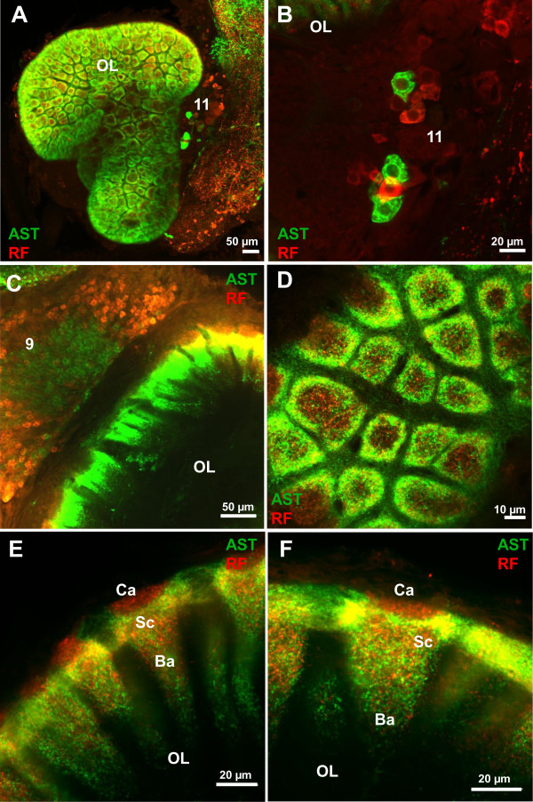
Figure 6.
Details of ASTir (green) and RFamide-like immunoreactivity (red) in the olfactory lobes (OL) as seen in confocal laser scanning microscopy. A the olfactory lobe in a projection of a series of confocal sections. B and C show two different types of local interneurons (note the spatial separation of AST and RFir somata in cluster 9 on C). D transverse and E, F longitudinal views of the glomeruli; for details see text. Abbreviations: 9, 11 cell cluster (9) and (11) of local interneurons, Ba base, Ca cap and Sc subcup of glomerulus, OL olfactory lobe.

