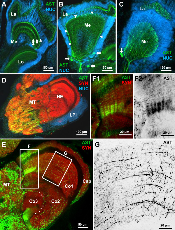
Figure 8.
Details of ASTir in the eyestalk neuropils AST immunoractivity.A,B,C: higher magnifications (conventional fluorescence with Apotome structured illumination), double labelling for ASTir (green) and the nuclear marker (blue). ASTir material in the medulla is arranged in three parallel layers, labeled by single, double, and triple arrowheads in A. Arrowheads in B indicate the outer (distal) layer of ASTir neurites of the medulla. Arrows in B and C identify ASTir somata. D higher magnification of the lateral protocerebrum (triple labelling, conventional fluorescence with Apotome structured illumination). E confocal-laser scan microscopic images (double labelling) of the hemiellipsoid body to show its subdivisions. Dashed line indicates the border of medulla terminalis and hemiellipsoid body. The boxed areas are magnified in F and G.

