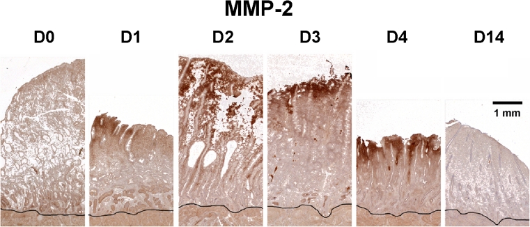
Fig. 6.
Immunohistochemistry of MMP-2 during the proliferative phase. Endometrial sections were stained for MMP-2 on days (D) 0,1,2,3,4 and 14 after P withdrawal. The black line marks the myometrial border. Strong dark brown staining for MMP-2 protein was confined to the upper functionalis zone during the menstrual phase (D1-4). The staining became nondetectable by day 14. Scale bar = 1 mm; applies to all images

