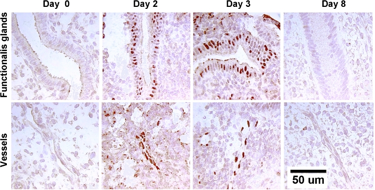
Fig. 7.
Immunohistochemistry of hypoxia inducible factor in the macaque endometrium. Endometrial sections were stained for hypoxia inducible factor protein on days 0, 2,3, and 8 after P withdrawal. A mouse monoclonal antibody against HIF-1α at a concentration of 1:1000 (Novus Biologicals) was used. Nuclear HIF-1α staining was low on day 0, increased strikingly on days 1–3 of the cycle and then declined to undetectable by day 8. These increases occurred primarily in the glands and the small blood vessels of the upper functionalis zone, presumably in response to local hypoxia. Scale bar = 50 um; applies to all images

