Figure 2.
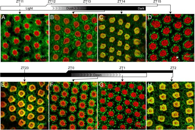
Light drives Aaop1 from the fused rhabdom to the cytoplasm. A–C, Mosquitoes were transitioned from light to dark conditions at ZT12. Lighting during the day was 100 lux and dimmed from 100 to 0 lux in the 1 h period from ZT12 to ZT13. Retinas were imaged for Aaop1 (green) and actin (red). Images show retinas dissected in the light 1 h before dusk (A), and 30 min (B) and 60 min (C) after complete darkness. D, Mosquitoes were retained in light conditions through the dusk time period and the retina was imaged for Aaop1 (green) and actin (red) at ZT15. The arrow marks one of the Aaop1-containing vesicles within the cytoplasm of an R8 photoreceptor. E–G, Mosquitoes were transitioned from dark to light conditions at ZT0. Whole-mount retinas were imaged for Aaop1 (green) and actin (red) 1 h before the lights gradually increase to 100 lux at dawn (E), and 30 min (F) and 60 min (G) after the dawn period. H, Mosquitoes were retained in dark conditions through the dawn time period and a retina imaged for Aaop1 (green) and actin (red) at ZT2, an hour after the time point shown in G for a mosquito subjected to the dawn conditions.

