Figure 5.

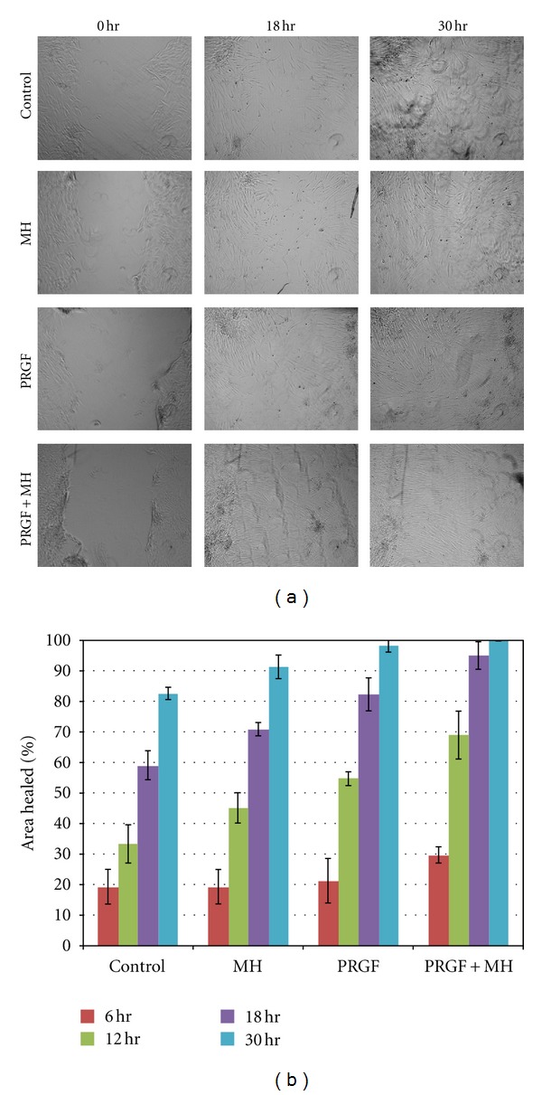
Light microscopy images of the in vitro wound healing assay taken at time 0, 18, and 30 hr after wounding (a). Graph of the mean % area healed for each of the test media at 6, 12, 18, and 30 hr after wounding (b).

Light microscopy images of the in vitro wound healing assay taken at time 0, 18, and 30 hr after wounding (a). Graph of the mean % area healed for each of the test media at 6, 12, 18, and 30 hr after wounding (b).